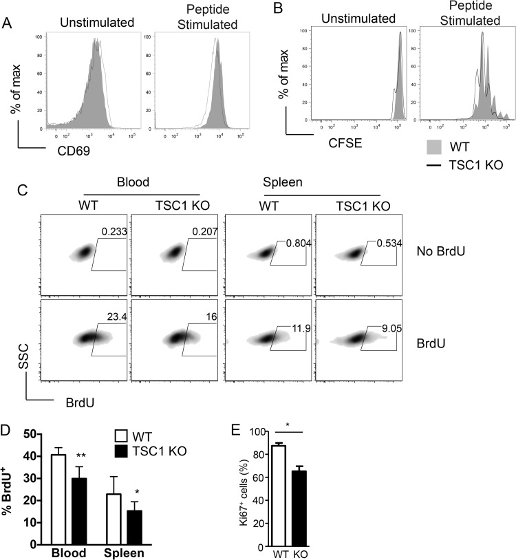
FIG 5.
Defective antigen-driven proliferation of TSC1-deficient CD8 cells in vivo. (A) Representative histograms of CD69 expression on WT and TSC1 KO OT1 cells that were cultured overnight with APCs that were not loaded or loaded with SIINFEKL peptide. (B) Representative histograms showing the CFSE dilutions among WT and TSC1 KO OT1 cells that were cultured for 65 to 72 h with APCs that were not loaded or loaded with SIINFEKL peptide. (C) Representative density plots showing BrdU incorporation in WT and TSC1 KO OT1 cells in the peripheral blood and spleen. Following competitive adoptive transfers, mice were injected with BrdU on day 5 postinfection and tissues were harvested after 16 h for staining and flow cytometric analysis. SSC, side scatter. (D) Percentages of BrdU+ cells among WT OT1 and TSC1 KO OT1 populations in the peripheral blood and spleen. The mean ± the standard error of the mean was calculated for four mice per group. (E) Percentages of Ki67+ donor-derived OT1 T cells 4 days after Lm-Ova infection. The data shown are representative of two or three independent experiments. *, P < 0.05; **, P < 0.01 (Student t test).

