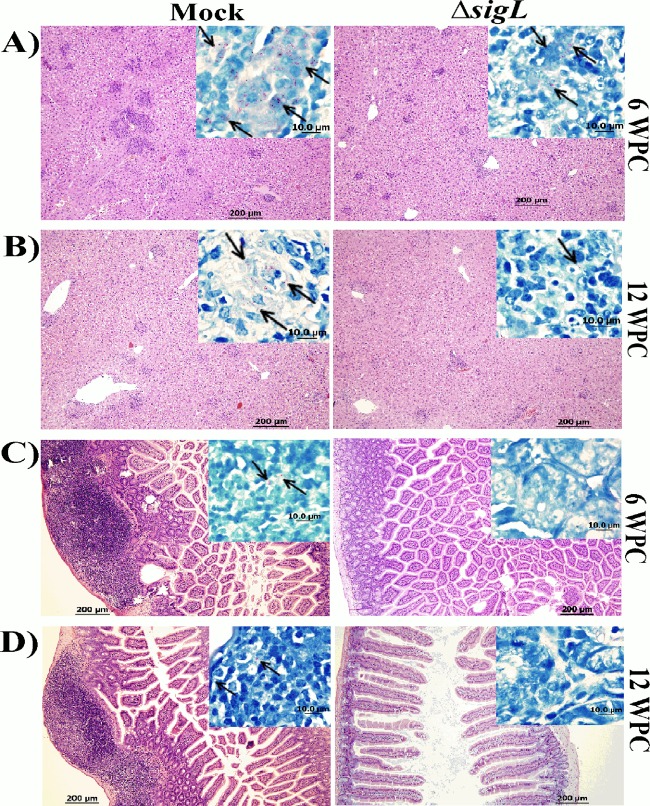
FIG 7.
Pathological analysis of mouse organs following vaccination. Photographs shows hematoxylin and eosin staining of liver (A and B) and intestine (C and D) sections from mock- and M. avium subsp. paratuberculosis ΔsigL-vaccinated animals following challenge with wild-type M. avium subsp. paratuberculosis at 6 WPC and 12 WPC. Magnification, ×100; bar, 200 μm. (Insets) Ziehl-Neelsen staining of both liver and intestine displayed more acid-fast bacilli in the mock-vaccinated animals than in the ones that received M. avium subsp. paratuberculosis ΔsigL vaccination. Magnification, ×1,000. Bar, 10 μm.

