FIG 7.
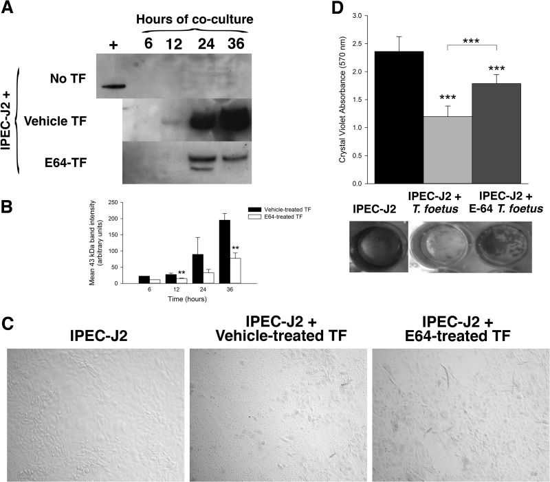
Cysteine protease activity mediates T. foetus cytopathogenicity. T. foetus was pretreated with the cysteine protease inhibitor E64 (300 μM) or vehicle (dH20) prior to coculture with IPEC-J2 monolayers for 6, 12, 24, or 36 h. (A) Immunoblot of uninfected IPEC-J2 monolayers and monolayers following 36 h of coculture with vehicle-pretreated T. foetus or E64-pretreated T. foetus for the presence of the M30 antigen of cleaved cytokeratin 18. (B) Densitometric analysis of the 3 immunoblots shown in panel A. **, P < 0.01 compared to vehicle-treated T. foetus (one-way ANOVA and post hoc Holm-Sidak test). Data represent n = 3 cultures per treatment and are reported as means ± SD. (C) Representative light microscopy images of uninfected IPEC-J2 monolayers and monolayers following 36 h of coculture with vehicle-treated T. foetus and E64-treated T. foetus (×40 magnification). (D) Spectrophotometric analysis of crystal violet absorbance of IPEC-J2 monolayers following coculture with vehicle-treated T. foetus or E64-treated T. foetus (A isolate) for 36 h. Representative wells are shown below treatment group columns. ***, P < 0.001 for vehicle-treated T. foetus compared to E64-treated T. foetus and compared to uninfected IPEC-J2 monolayers (one-way ANOVA and post hoc Holm-Sidak test). Data represent n = 6 cultures per treatment group and are representative of 3 different feline T. foetus isolates. Data are reported as means ± SD. Data reported from uninfected and T. foetus-infected monolayers are also shown in Fig. 3.

