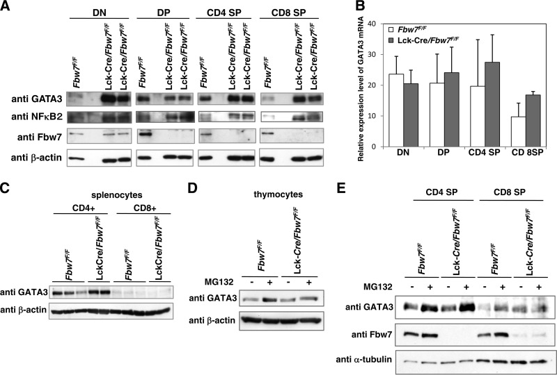
FIG 3.
Loss of Fbw7 stabilizes GATA3 protein in mouse thymocytes. (A) Representative immunoblot analysis of Fbw7 and its target proteins in the subsets of thymocytes from Fbw7flox/flox or Lck-Cre/Fbw7flox/flox mice. DN, DP, CD4 SP, and CD8 SP cells were purified from Fbw7flox/flox or Lck-Cre/Fbw7flox/flox mice at 8 weeks of age using flow cytometry and lysed. The lysates were subjected to immunoblot analysis with the indicated antibodies. (B) qRT-PCR analysis of GATA3 expression in the sorted FACS fractions obtained in panel A. The amount of transcripts was normalized against that of GAPDH as an internal standard. Data are means ± SD of values from three Fbw7flox/flox and three Lck-Cre/Fbw7flox/flox mice. (C) Purification of CD8+ and CD4+ T cells from mouse splenocytes from Fbw7flox/flox or Lck-Cre/Fbw7flox/flox mice at 8 to 9 weeks of age was performed with CD8+ positive selection and CD4+ negative selection by the magnetic bead method, respectively. Immunoblot analysis of whole lysates from each purified splenic T-cell subset was performed with the indicated antibodies. (D) T-cell subsets obtained from Fbw7flox/flox or Lck-Cre/Fbw7flox/flox mice at 8 weeks of age were cultured in RPMI 1640 medium for 2 h and incubated with 20 μM MG132 for 4 h. Cells were lysed and subjected to immunoblot analysis with the indicated antibodies. (E) Whole thymocytes obtained from Fbw7flox/flox or Lck-Cre/Fbw7flox/flox mice at 8 weeks of age were cultured in RPMI 1640 medium for 4 h and incubated with 20 μM MG132 for 5 h. Cells were lysed and subjected to immunoblot analysis with the indicated antibodies.

