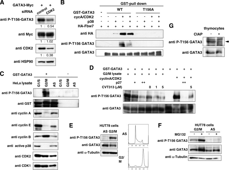
FIG 8.
Phosphorylation at Thr-156 in GATA3 by CDK2 is required for association of Fbw7 and is executed in HUT78 cells during G2/M phase and in thymocytes of mice. (A) Depletion of CDK2 reduced phosphorylation of Thr-156 in GATA3 in HEK293 cells. HEK293 cells were transfected with a WT GATA3 expression plasmid and CDK2 or a control siRNA. After 43 h of transfection, cells were treated with okadaic acid and MG132 for 5 h. Cell lysates were prepared and subjected to immunoblot analysis with the indicated antibodies. The numbers reflect the ratio of the levels of the indicated proteins between the CDK2 siRNA-transfected cells and control cells. (B) GATA3 binding to Fbw7 in vitro is Thr-156 phosphorylation dependent. Purified WT or T156A GST-GATA3 was incubated with the indicated kinases in reaction buffer at 30°C for 30 min. Lysates from HEK293 cells transfected with HA-Fbw7 were immunoprecipitated with HA antibody, and the immunocomplexes containing Fbw7 were incubated with phosphorylated GST-GATA3 in vitro as indicated. To analyze GATA3 and Fbw7 binding, the GST fusion protein complexes were precipitated using glutathione-Sepharose beads and subjected to immunoblotting with the indicated antibodies. (C and D) Phosphorylation of recombinant GATA3 in vitro. Reaction products were then subjected to immunoblot analysis with the indicated antibodies. (C) Phosphorylation of Thr-156 in GATA3 in G2/M-arrested cells. GST-GATA3 was incubated with lysate prepared from either cells arrested in G1/S or G2/M phase or nonsynchronized (AS) HeLa cells. (D) Effects of CDK2 inhibition on Thr-156 phosphorylation of GATA3 in G2/M cell lysate. The responsible kinase in G2/M-phase cell lysate is CDK2. GST-GATA3 was incubated with the indicated kinase sources in the absence or presence of CDK2 inhibitor (CVT313) or competitor (p27). (E and F) Thr-156 phosphorylation of endogenous GATA3 in G2/M phase in T-cell lymphoma HUT78 cells. G2/M-arrested and asynchronous (AS) HUT78 cells were prepared as indicated in Materials and Methods. Cell lysates were prepared and subjected to immunoblot analysis with the indicated antibodies. (F) G2/M-arrested and asynchronous cells were treated with or without MG132 for 5 h before harvest. Cell lysates were prepared and subjected to immunoblot analysis with the indicated antibodies. (G) Cell lysate from whole thymocytes obtained from an Fbw7flox/flox mouse at 6 weeks of age was incubated with or without calf intestinal alkaline phosphatase (CIAP) at 37°C for 30 min and subjected to immunoblot analysis with the indicated antibodies.

