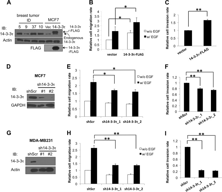
FIG 1.
14-3-3τ regulates breast cancer cell migration and invasion. (A) A stable MCF7 cell line expressing 14-3-3τ–FLAG or an empty vector (Vec) was established, and lysates were subjected to SDS-PAGE analysis side by side with four primary human breast tumor samples (5T, 37T, and 10T are from the 14-3-3τ overexpression group, whereas 9T is from the nonoverexpression group [3]). Total cell lysates were immunoblotted (IB) using anti-FLAG and anti-14-3-3τ antibodies, respectively, to verify the expression of 14-3-3τ–FLAG. The expression of β-actin serves as a loading control. (B) Overexpression of 14-3-3τ increased MCF7 cell migration. MCF7 stable cell lines were subjected to a Transwell cell migration assay. EGF was added to the lower chamber of Transwells, and cells were allowed to migrate for 6 h. The relative migration rate was defined as the fold increase of migrated cells compared to the level for unstimulated vector control cells. Data shown are the means ± standard errors of the means from four independent experiments. *, P < 0.05 (two-tailed t test). w/, with; w/o, without. (C) 14-3-3τ-overexpressing MCF7 cells had a higher invasion rate. Stable MCF7 cells were starved in serum-free DMEM for 24 h and then seeded on the upper chamber of a Matrigel-coated insert. Five percent serum-containing medium was added to the lower chamber, and cells were allowed to invade for 24 h. **, P < 0.001 (n = 3) (two-tailed t test). (D) MCF7 cells were infected with lentiviruses expressing either control scrambled shRNA (shScr) or one of the 14-3-3τ shRNAs (1 and 2) and selected with puromycin. Cell lysates were subjected to immunoblotting to confirm the knockdown of 14-3-3τ. (E) 14-3-3τ knockdown attenuated MCF7 cell migration rate. Stable MCF7 cell lines expressing an shScr or 14-3-3τ shRNAs were subjected to a Transwell cell migration assay in the absence or presence of EGF (0.1 μg/ml for 5 h) as described for panel B. *, P < 0.05 (n = 3) (two-tailed t test). (F) Cell invasion was reduced upon 14-3-3τ depletion. Stable MCF7 cell lines expressing an shScr or 14-3-3τ shRNAs were subjected to a Matrigel invasion assay as described for panel C. **, P < 0.001 (n = 4) (two-tailed t test). (G) MDA-MB231 cells were infected with lentiviruses expressing either control shScr or 14-3-3τ shRNA (1 and 2) and selected with puromycin. Cell lysates were subjected to immunoblotting to confirm the knockdown of 14-3-3τ. (H) MDA-MB231 cell lines expressing a scrambled shRNA or 14-3-3τ shRNAs were subjected to a Transwell cell migration assay in the absence or presence of EGF (0.01 μg/ml for 5 h). **, P < 0.001 (n = 3) (two-tailed t test). (I) 14-3-3τ-depleted MDA-MB231 cell lines were subjected to a Matrigel invasion assay as described for panel C. **, P < 0.001 (n = 3) (two-tailed t test).

