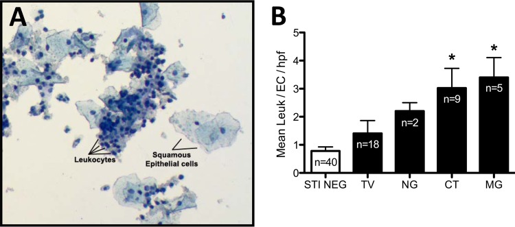
FIG 2.
Quantification of cervical leukocytes from liquid cytology specimens. ThinPrep PreservCyt specimens from low-risk Louisiana women were screened for M. genitalium using the TaqMan PCR system described herein, as well as for C. trachomatis, N. gonorrhoeae, and T. vaginalis. All subjects with coinfections among those positive for M. genitalium, C. trachomatis, N. gonorrhoeae, and T. vaginalis were excluded from the calculation of leukocyte modulation. Those specimens that were monopositive for each STI and a randomly selected subset of specimens negative for all tested STIs (n = 40) were Diff-Quik stained, followed by quantification of cervical epithelial cells and leukocytes. (A) Representative microscope field from a stained ThinPrep PreservCyt specimen illustrating squamous epithelial cells and cervical leukocytes. Differing ratios of leukocytes to epithelial cells are readily observed using the preparation and staining paradigm. (B) Comparison of cervical leukocyte counts among women with and without STIs presented as the ratio of leukocytes to epithelial cells per high-powered field (HPF) compiled from five representative microscope fields. Significant differences between leukocyte/epithelial cell ratios among subjects are indicated with an asterisk (P < 0.05; ANOVA).

