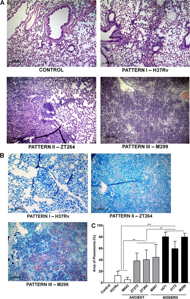
FIG 5.
Histopathological changes in the lungs of M. tuberculosis-infected mice. The lungs were obtained from infected mice as described in the legend to Fig. 3. (A) Representative hematoxylin-and-eosin-stained lung sections, demonstrating three main pathological patterns. Bars, 200 μm. (B) Representative lung sections stained by Ziehl-Neelsen method. In pattern I, induced by the strains H37Rv and M442, small and medium-sized peribronchial and perivascular granulomas, consisting of macrophages and lymphocytes, are observed. Small numbers of intracellular bacteria are seen. In pattern II, induced by strains zt264, zt272, and M467, moderate, multifocal granulomatous pneumonia is observed. Increasing numbers of intracellular bacteria are seen. In pattern III, induced by strains M299, 2172, and 1471, extensive diffuse granulomatous pneumonia with areas of necrosis and alveolitis is observed. Large numbers of intracellular and extracellular bacteria are seen. Bars, 200 μm. (C) Morphometric analysis of the inflammatory lung area. Results of two experiments (three mice in each group in each experiment). Data represent means plus SD. Significant differences were determined between the groups of pattern III strains (black columns), pattern II strains (gray columns), and pattern I strains (white columns) and indicated by asterisks as follows: **, P < 0.01; ***, P < 0.001.

