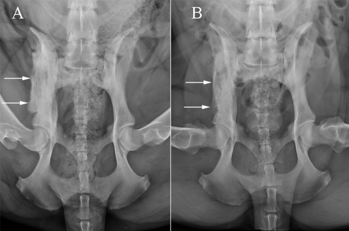
FIG 1.

Ventrodorsal projections of the pelvis at presentation (A) and 3 months later (B). There is extensive mixed periosteal proliferation and permeative lysis along the right ilial body and wing in the initial radiograph, which are smoother and less severe in the subsequent image.
