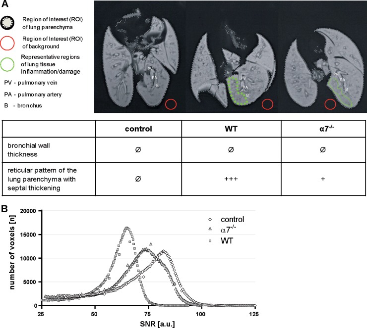
FIG 4.
Total-lung imaging of WT and importin-α7−/− mice upon influenza virus infection. Macroscopic lung tissue damage was analyzed ex vivo by MRI. Control mice treated with PBS were compared with WT and α7−/− mice infected with 106 PFU of PR8:NS1-GFP virus. Lungs were processed at day 3 p.i. (A) T2-weighted MRI presents virus-induced lung inflammation and damage with the formation of a hypointense reticular pattern within the lung parenchyma, which was most severe in WT mice, followed by α7−/− mice. In contrast, uninfected control mice showed homogeneous lung tissues. (B) According to this qualitative rating, the quantitative distribution analysis of the SNR of each voxel of the lungs demonstrated the highest values in control mice (white diamonds) and reduced SNRs in α7−/− (light gray triangles) and WT mice (dark gray squares). a.u., arbitrary units. See also Videos S1, S2, and S3 in the supplemental material.

