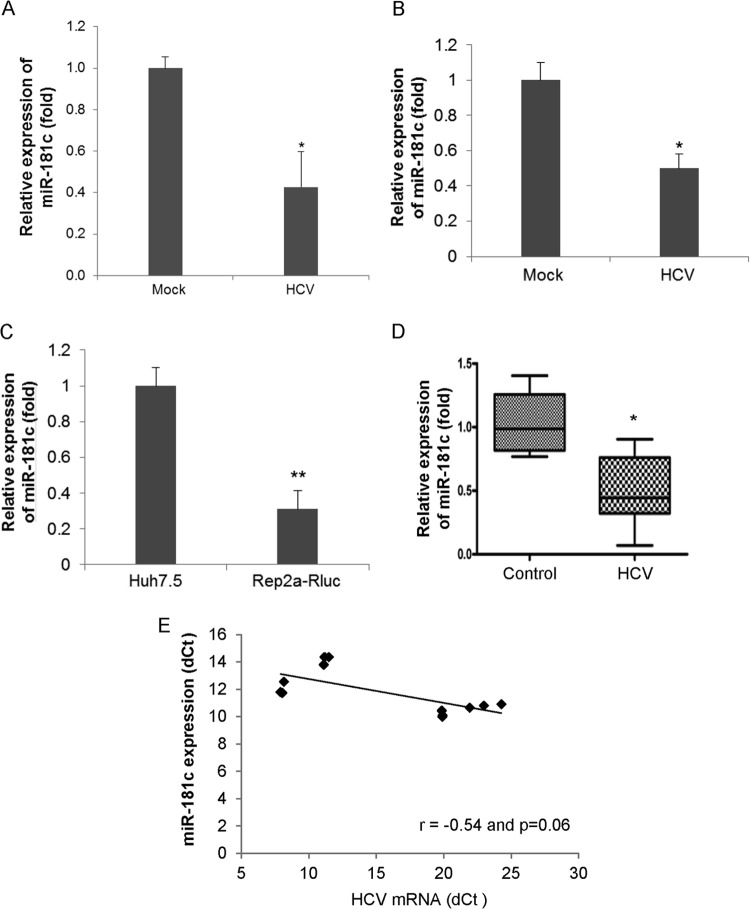
FIG 1.
HCV infection suppresses miR-181c expression. (A) Primary human hepatocytes were mock treated or HCV infected for 96 h. Total RNA from mock- or virus-infected cells was extracted, and the expression level of miR-181c was measured by qRT-PCR and normalized to the expression level of U6. (B) Huh7.5 cells were mock treated or HCV infected (multiplicity of infection of 0.5) for 72 h. Total RNA from mock- or virus-infected cells was extracted, and the expression level of miR-181c was measured and normalized to the expression level of U6. (C) Total RNA was extracted from Huh7.5 cells as a control or Huh7.5 cells harboring the HCV genome-length replicon (Rep2a-Rluc), and the expression level of miR-181c was determined by qRT-PCR. Data are presented as the means and standard deviations from three independent experiments. (D) Total RNA was extracted from control (not infected with HCV or HBV) and HCV-infected liver biopsy specimens. miR-181c expression was analyzed by real-time PCR, using U6 RNA as an internal control. A comparison of miR-181c levels in control and HCV-infected (n = 6) liver tissues is shown. *, P < 0.05; **, P < 0.01. (E) Correlation plot for miR-181c expression versus HCV RNA levels in liver biopsy specimens. We observed a strong inverse correlation between miR-181c and intrahepatic HCV RNA levels (r = −0.54; P = 0.06). Statistical analysis was done by using the Spearman correlation.

