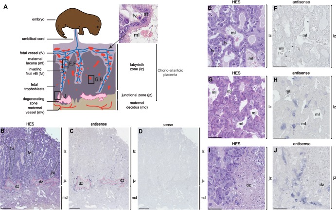
FIG 8.
Structure of the placenta and in situ hybridization for syncytin-Mar1 expression on woodchuck placental sections. (A) Schematic representation (based on panel B) of the woodchuck placenta with, from fetus to mother, the labyrinthine zona (lz), the junctional zone (jz), and the maternal decidua (md). An enlarged view of a fetal villus tissue section stained with hematoxylin-eosin-saffron (HES) (upper right) shows the multinucleated syncytiotrophoblast (st) generated by fusion from the underlying mononucleated cytotrophoblast cells forming the interface between the fetal vessel (fv) and the maternal lacuna (ml). (B to J) Sections of placenta from woodchuck at midgestation, with the positions of the E to J panels indicated in panel A. (B, E, G, and I) HES staining of placental sections with the three layers (lz, jz, and md) indicated; in panel B the fetal villi (fvi) are delineated with a black dotted line, and the degenerating zone (dz) with a pink dotted line. (C, D, F, H, and J) In situ hybridization on serial sections observed at different magnifications, using digoxigenin-labeled antisense (C, F, H, and J) or sense (negative control [D]) riboprobes revealed with an alkaline phosphatase-conjugated anti-digoxigenin antibody. (E and F) Intermediate magnification of the labyrinth zone in the fetal villi delineated in panel A, with no detectable labeling. (G and H) Intermediate magnification of the labyrinth zone in formation delineated in panel A, with labeling close to the maternal lacunae. (I and J) Intermediate magnification of the junctional zone delineated in panel A, with labeling abutting the degenerating zone on the right. Scale bars: 0.5 mm for the serial sections (B to D), 50 μm for panels E and F, and 100 μm for panels G to J.

