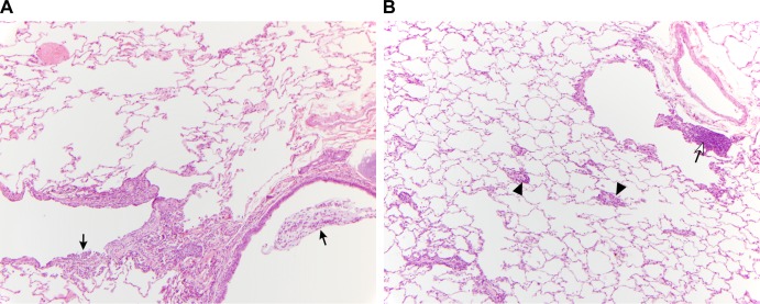
FIG 3.
Comparison of lung pathology results 2 days postchallenge with H5N1 wt virus in AGMs immunized i.n. with a sprayer versus i.n.-plus-i.t. immunization. (A) Section of lung from an intranasally (sprayer) vaccinated monkey (AGM7). There are low to moderate numbers of neutrophils and macrophages visible adjacent to a bronchus (lower right) and bronchiole (lower left). Black arrows indicate neutrophils within the lumen of these airways. Perivascular lymphocyte cuffing is absent. (B) Section of lung from an i.n.-plus-i.t.-vaccinated monkey (AGM8). Arrowheads indicate small-caliber blood vessels surrounded by low to moderate numbers of lymphocytes (perivascular lymphocyte cuffing). The open arrow indicates aggregated lymphocytes adjacent to a bronchiole (lymphocyte hyperplasia).

