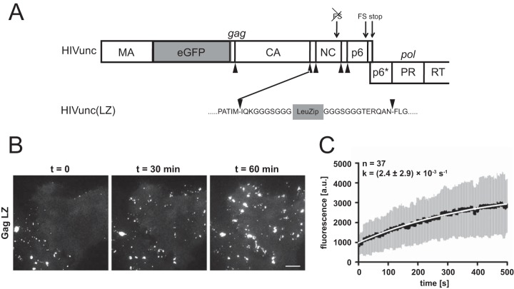
FIG 3.
Assembly kinetics of a LZ-carrying HIV derivative. (A) Scheme of the Gag/Pol-encoding region of pCHIVunceGFP illustrating the insertion site for the leucine zipper. Arrowheads indicate cleavage sites for the HIV-1 protease. The leucine zipper sequence is flanked by short G/S-rich linker sequences. FS, translational frameshift signal; RT, reverse transcriptase. (B) Assembly of HIVunc(LZ)eGFP at the plasma membrane of a particle-producing cell. HeLa cells were transfected with an equimolar mixture of pCHIVunc(LZ) and pCHIVunc(LZ)eGFP. At 20 h after transfection, cells were analyzed by live-cell TIR-FM as described in Materials and Methods. Shown are individual frames from Movie S2 in the supplemental material recorded at the indicated times. Bar, 10 μm. (C) Rate of Gag assembly determined for HIVunc(LZ)eGFP. HeLa cells were transfected with equimolar mixtures of pCHIVunc(LZ) and pCHIVunc(LZ)eGFP. At 20 h after transfection, cells were analyzed by TIR-FM (see Movie S2 in the supplemental material), and individual assembly sites were tracked. Fluorescence intensities from the exponential assembly phase from 37 individual sites from 2 experiments were averaged as described previously (38). The graphs show mean values (black line) and standard deviations (gray bars); mean values were fitted to a saturating single exponential equation (white line), yielding the indicated rate constant. a.u., arbitrary units.

