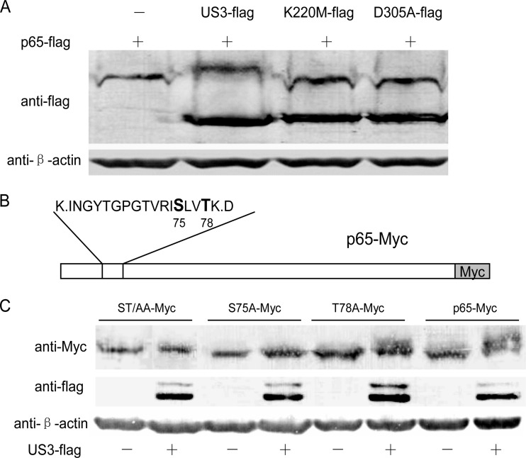
FIG 6.
US3 hyperphosphorylates p65. (A) HEK293T cells were transfected with pCMV-p65-Flag alone or with pCMV-US3-Flag, pCMV-K220M-Flag, and pCMV-D305A-Flag separately. WBs were performed with anti-Flag and anti-β-actin monoclonal antibody. (B) Schematic diagram of the potential hyperphosphorylation sites (S75 and T78) of p65. (C) p65 and its three mutants (ST/AA-Myc, S75A-Myc, and T78A-Myc) were cotransfected with or without US3-Flag. Thirty-six hours posttransfection, cell lysates were subjected to WB analysis.

