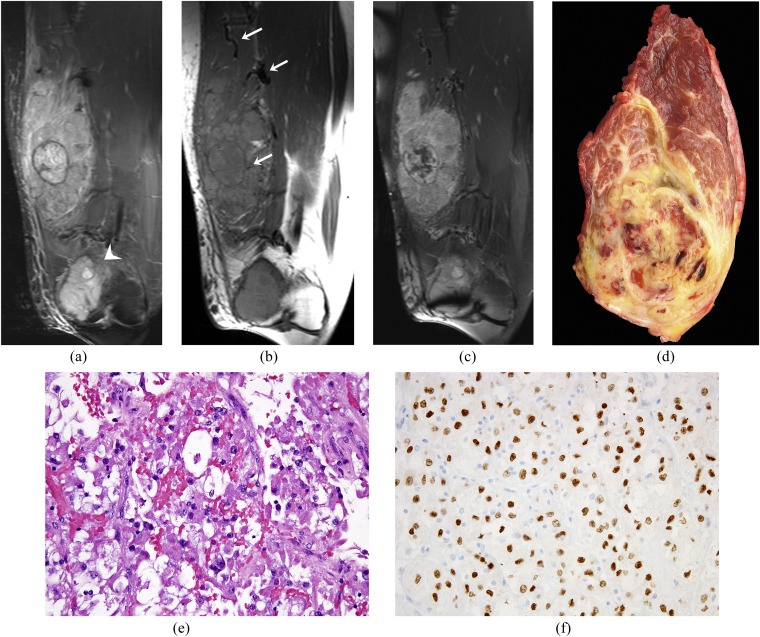
Figure 1.
Images of a 29-year-old male with alveolar soft part sarcoma (ASPS) of the right thigh. (a–c) Coronal short tau inversion–recovery, pre- and post-gadolinium T1 weighted fat-suppressed MR images reveal a heterogeneous T1 and T2 hyperintense (relative to the adjacent muscles) mass involving the extensor compartment of the thigh in relation to the vastus lateralis with intense post-gadolinium enhancement. Note the prominent feeder vessels and flow voids (arrows, b). A metastatic lesion involving the distal femur is also visualized (arrowhead, a). Both the lesions were resected. (d) Gross resected specimen of the thigh sarcoma shows extensive necrosis. (e) On histopathology, there is a nested architecture, with the tumour being composed of large epithelioid cells with voluminous cytoplasm. (f) By immunohistochemistry, the tumour cells show strong nuclear staining for TFE3, reflecting the presence of a t(X; 17) translocation with the ASPSCR1–TFE3 fusion.

