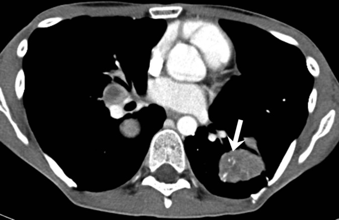
Figure 2.

Image of an 18-year-old male with ASPS metastatic to the lungs. Axial contrast-enhanced CT image of the chest shows bilateral hyperattenuating pulmonary metastases. The left lower lobe lesion shows linear enhancing intratumoral vessels (arrow).
