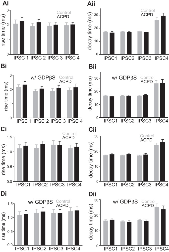
Fig. 2.
Effects of ACPD on the rise time (20–80%) and the decay time (80–20%) of the 4 evoked IPSCs in V1. A: effects of ACPD on rise time and decay time of IPSCs recorded in layers 2/3 cells while stimulating in layer 4 in V1. Ai: rise time of the 4 IPSCs before (Control) and after ACPD application (ACPD) using normal intracellular solution. ACPD application had no effect on the rise time of any of the 4 IPSCs. Aii: decay time of the 4 IPSCs before (Control) and after ACPD application (ACPD). ACPD application had no effect on the decay time of any of the 4 IPSCs. B: effects of ACPD on rise time and decay time of IPSCs recorded in layers 2/3 cells while stimulating in layer 4 in V1 with GDPβS in the intracellular solution. ACPD application produced no significant change in the rise time or the decay time of the IPSCs. C: effects of ACPD on rise time and decay time of IPSCs recorded in layer 4 cells while stimulating in an adjacent location within layer 4 in V1 using normal intracellular solution. ACPD application had no significant change in the rise time or the decay time of IPSCs. D: no significant effect of ACPD application on the rise time or the decay time of IPSCs recorded in layer 4 cells while stimulating in an adjacent location within layer 4 in V1 with GDPβS in the intracellular solution.

