
Figure 3.
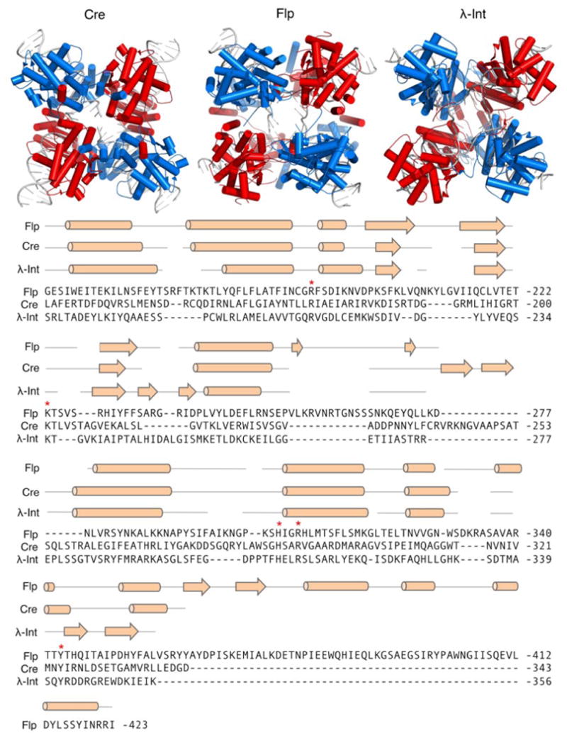
Overview of the tyrosine recombinases. Top: Structures of the Cre, Flp, and λ-Int tetramers complexed with their respective DNA targets. Pairs of recombinase dimers are colored red and blue. DNA depicted as gray cartoon. PDB IDs are Cre: 1CRX (Guo et al., 1997), Flp: 1FLO (Chen et al., 2000), and λ-Int: 1Z1G (Biswas et al., 2005). Bottom: Sequence alignment of Cre, Flp, and λ-Int C-terminal domains. Secondary structural elements for each enzyme are indicated above alignment. Cylinders and arrows indicate α-helix and β-sheet secondary structures, respectively. Red asterisks indicate conserved amino acid residues critical for catalysis.