
Figure 4.
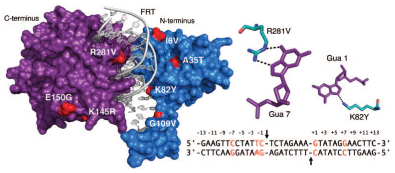
Distribution of mutations that contribute to evolved Flp specificity. Left: Surface illustration of the Flp monomer bound to FRT DNA target. Mutations that contribute to the interactions between evolved Flp variants and unnatural FRT targets (Voziyanov et al., 2003) are highlighted red. DNA depicted as a gray cartoon. Flp N-terminal domain colored blue, C-terminal domain colored purple. Not visible are the substitutions V266A and N264I. PDB ID is 1FLO (Chen et al., 2000). Right upper: The interactions between Arg 281 of wild-type Flp and Gua 7 of wild-type FRT, and Lys 82 and Gua 1 are shown. Gua 7 and Gua 1 were among five positions substituted in the mutant FRT site by Voziyanov et al. (Right lower) FRT target. Positions substituted for evolution studies highlighted red. Black arrows indicate the location of the scissile phosphates.