
Figure 6.
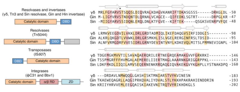
Overview of the serine recombinases. Left: Domain organization of the serine recombinases. α/β RD and ZD indicate C-terminal α/β recombinase and zinc-nucleated integrase domains, respectively (Rutherford et al., 2013). Gray indicates domains of unknown function. Right: Comparison of representative members of the resolvase/invertase family of serine recombinases. Conserved residues are shaded pink. Secondary structural elements within the γδ resolvase are indicated above alignment. Cylinders and arrows indicate α-helix and β-sheet structures, respectively. Asterisk indicates the conserved serine residue critical for catalysis.