
Figure 10.
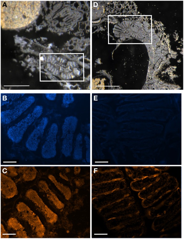
Digestive ceca of symbiotic vs. symbiont-deprived 5th instar Sibaria englemani nymphs. (A–C) Light microscopy, DAPI, and FISH microscopy, respectively, of the ceca of a symbiotic nymph, showing dense bacteria within the crypt spaces. (A) shows the non-symbiont containing sac-like midgut region (M3), as described in Futahashi et al. (2013), at top left (D–F) Light microscopy, DAPI, and FISH microscopy, respectively, of the ceca of a symbiont-deprived nymph, showing lack of cecal bacteria and visual differences in gross ultrastructure. (A,D) Light images of sections through the midgut ceca, embedded in, and then removed from, Steedman's wax prior to hybridization. Boxes denote areas of subsequent DAPI and fluorescent 16S rRNA probing. (B,E) Fluorescent images showing boxed regions in (A,D) counter-stained with DAPI, observed in blue. (C,F) Fluorescence in situ hybridization (FISH) microscopy of the boxed regions in (A,D), hybridized with the Eub338 probe set labeled with Cy3, shown in orange. Light image scales, 2 mm. Fluorescent image scales, 20 μm.