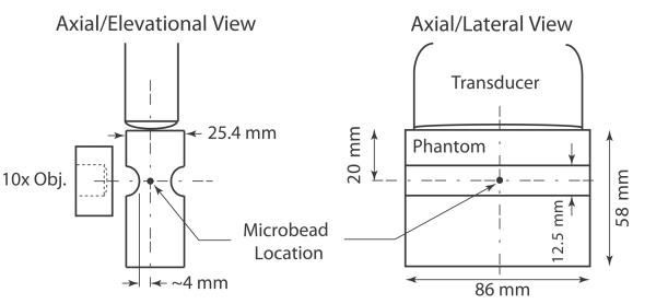
Figure 1.
Schematics of phantom design and confocal alignment of optical and acoustic foci showing the axial/elevational view and the axial/lateral view. Although only one microbead is shown in these schematics, many microbeads (~2.28 × 10⁷ beads) were dispersed throughout the whole volume of the phantom.

