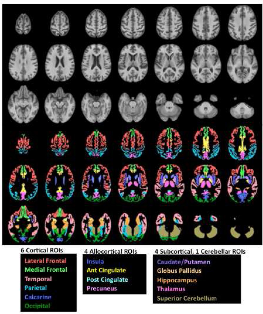
Figure 1.
Top panel in gray scale: Axial slices from the SRI24 atlas of the superior (top left) to inferior (bottom right) brain regions. Bottom panel in color: Six bilateral cortical and four allocortical gray matter regions and four subcortical tissue regions overlaid on the SRI24 atlas and color-coded by structure name. Ant, anterior; Glob, globus; Post, posterior; ROI, region of interest.

