Figure 2.
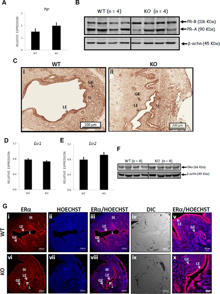
Pgr, Esr, PR, and ER are expressed at similar levels in the uteri of Kiss1−/− and WT mice. Relative to the expression of two housekeeping genes (Hprt, Sdha), the expression of Pgr, Esr1, and Esr2 (A, D, and E) was determined by quantitative RT-PCR [for superovulated WT female mice, n = 6; for E2/gonadotropin-primed/superovulated KO (Kiss1−/−) mice, n = 4]. The spatial distribution and expression levels of PR-A and PR-B (the PR antibody detects both isoforms) were determined by immunohistochemistry, whereas that of ERα was determined by immunofluorescence (C and G) (for WT, n = 4; for KO (Kiss1−/−), n = 4). The expression level of PR-A, PR-B, and ERα was also determined by Western blotting (B and F) (for WT, n = 4; for knockout (KO) (Kiss1−/−), n = 4). Error bars represent SEM. DIC, differential interference contrast; GE, glandular epithelium; L, lumen; LE, luminal epithelium; St, stroma; HOECHST, nucleic acid stain that identifies nuclei.

