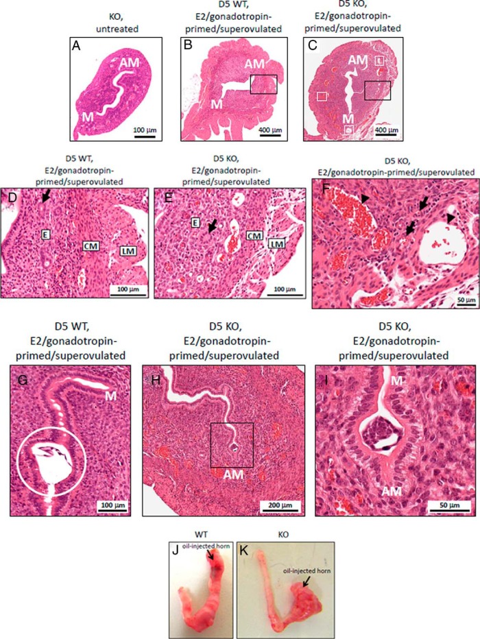
Figure 4.
Histology and morphology of Kiss1−/− and WT mice uteri. Anatomy of the representative uteri of the age-matched female mice is as follows: untreated KO (Kiss1−/−) female mice (A), E2/gonadotropin-primed/superovulated WT female mice (B), and E2/gonadotropin-primed/superovulated KO female mice (C) on day 5 after mating; white boxes show dilated blood vessels. Please note the different magnifications in panel A vs panels B and C. D–F, Higher-magnification images of the uterine sections corresponding to E2/gonadotropin-primed/superovulated WT and knockout (KO) female mice on day 5 after mating. D, Normal uterine architecture (corresponds to black outlined box in panel B) from E2/gonadotropin-primed/superovulated WT female mice on day 5 after mating. E, Abnormal uterine architecture (corresponds to black outlined box in panel C) from E2/gonadotropin-primed/superovulated KO female mice on day 5 after mating. F, Dilated vessels (corresponds to lower part of image shown in panel C) of E2/gonadotropin-primed/superovulated KO female mice on day 5 after mating. G, An implanted WT embryo (enclosed within white outlined circle) is observed near the antimesometrial (AM) pole of an E2/gonadotropin-primed/superovulated WT female mouse on day 5 after mating. H, Nonimplanted Kiss1+/− embryo (seen within black outlined box) is observed near the AM pole of an E2/gonadotropin-primed/superovulated KO female mouse on day 5 after mating. I, Nonimplanted Kiss1+/− embryo (as seen within black outlined box in panel H) is shown at higher-power magnification. Please note the different magnifications shown in panels G–I. J, Representative images of uterine horns after the injection of oil into the left horn of the hormone-primed ovariectomized WT (J) (n = 4) and KO female (K) (n = 4). CM, circular muscle of the myometrium; D, endometrium; GO, gland opening to uterine lumen; LM, longitudinal muscle; M, mesometrial pole.

