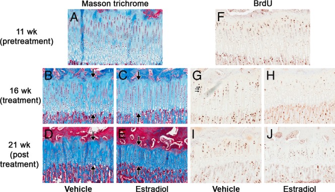
Figure 1.

PT growth plate morphology and proliferation before (11 weeks), at the end of treatment (16 weeks), and 5 weeks after the estrogen treatment was stopped (21 weeks). Representative sections of PT growth plates from 11-(A and F), 16- (B, C, G, and H), and 21-week-old (D, E, I, and J) control (A, B, D, F. G, and I) and estrogen-treated (C, E, H, and J) animals stained with Masson Trichrome (A–E) or by BrdU immunohistochemistry (F–J). Estrogen treatment (16-week time point) affected the structure of the growth plate, decreasing the overall height as well as the number of cells in each zone. Interestingly, 5 weeks after the estrogen treatment was stopped (21-week time point) these structural changes remained advanced. Estrogen treatment also decreased the number of BrdU-labeled cells in estrogen treated animals (H) compared with controls (G). However, 5 weeks after the estrogen treatment was stopped (21 weeks) the number of BrdU-labeled cells in estrogen- treated animals (J) was similar to that of controls (I). Large arrows indicate the height of the growth plate, whereas small arrows indicate BrdU-labeled cells.
