
Figure 4.
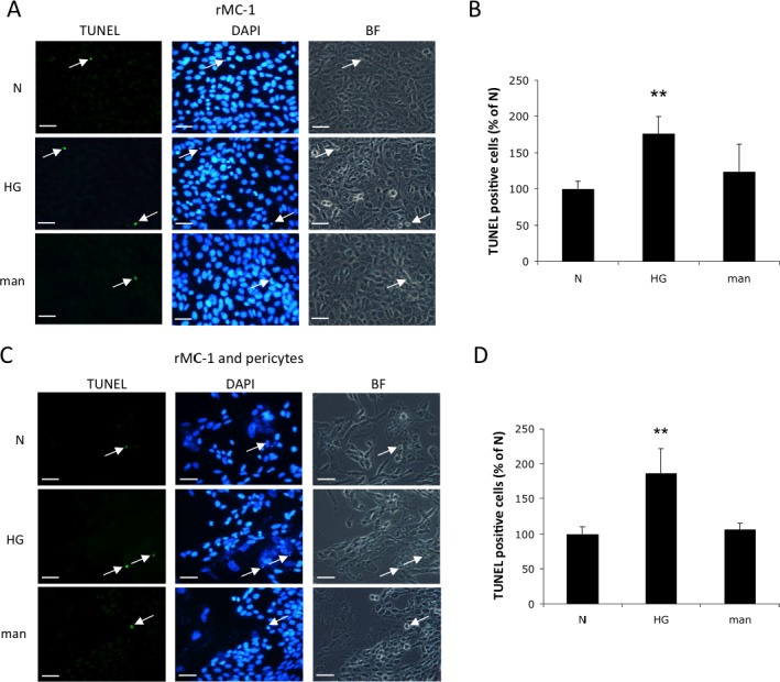
High glucose promotes apoptosis in retinal Müller cells and pericytes. (A) Representative images of TUNEL-positive cells (arrows) in rMC-1. Scale bars: 50 μm. (B) Graphical illustration of cumulative data showing TUNEL-positive cells were significantly increased in rMC-1 grown in HG medium compared with those grown in N or mannitol (man) medium. (C) Representative images of TUNEL-positive cells (arrows) in cocultures of rMC-1 and pericytes. Scale bars: 50 μm. (D) Graphical illustration of cumulative data showing TUNEL-positive cells were significantly increased in cocultures of rMC-1 and pericytes grown in HG medium compared with those grown in N or mannitol medium. Data are expressed as the mean ± SD. **P < 0.01. n = 4.