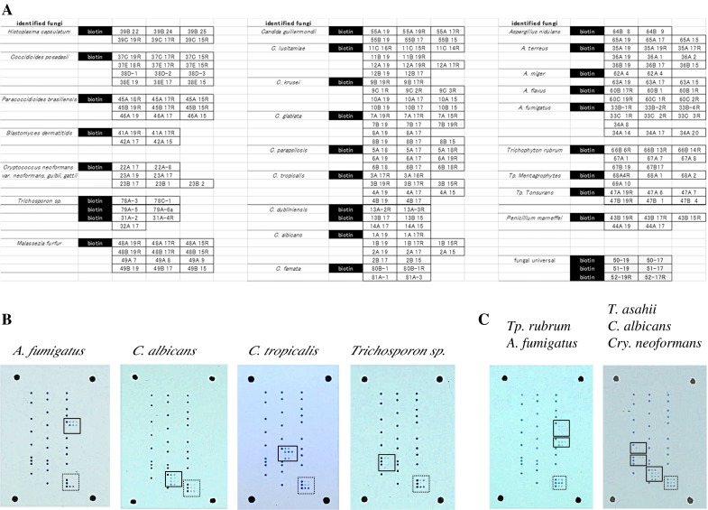
Fig. 1.

a Example layout of capture probes on microarray slide. Probe names correspond to probe names listed in Table 1. The black column labeled “biotin” indicates the spots for positive control (biotinylated-poly-T) and positional marker. b Typical hybridization patterns using fungal suspension of different fungal species as PCR template. Species-specific signals are enclosed in solid line frames, while universal signals for fungi are enclosed in dotted line frames. These figures show representative results for A. fumigatus, Trichosporon asahii, C. tropicalis, and C. albicans. c Simultaneous hybridization of several species in one array slide. Fungal cell mixtures of C. albicans, Cryptococcus neoformans, and T. asahii or of A. fumigatus and Trichophyton rubrum were directly used as template for PCR amplification and detected on the array slide
