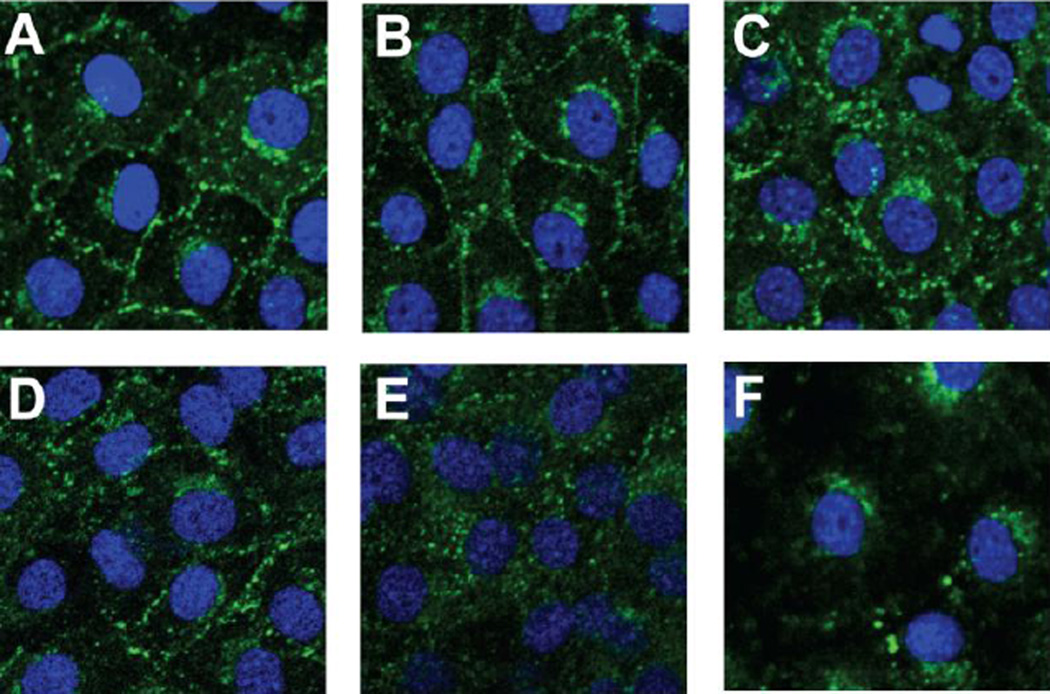
Figure 5.
Connexin 43 membrane localization is altered in BALBLpsd mice. Cx43 immunostaining in C10 cells exposed to BALF from BALB and BALBLpsd mice for 4 h. DAPI was used as the nuclear stain and Alexa-Fluor 488 linked to specific Cx43 antibodies. (A) C10 cells treated with serum-deprived media as the control; (B) C10 cells treated with BALF from BALB oil-treated mice; (C) C10 cells treated with BALF from BALB BHT-treated mice; (D) C10 cells treated with BALF from BALBLpsd oil-treated mice; (E) C10 cells treated with BALF from BALBLpsd BHT-treated mice; (F) C10 cells treated with TPA as a tumor promoter control to demonstrate removal of Cx43, as observed previously [16,29]. N=2–3 for each treatment group and repeated twice. Magnification, 1000×.

