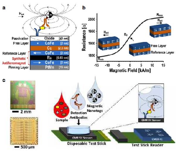
Figure 5. Giant magnetoresistance (GMR) detection of biomarkers.
(a) GMR sensors consist of alternating layers of ferromagnetic and non-magnetic materials. The magnetization of a reference layer is pinned and the magnetization of a free layer is able to change with an applied field. The presence of MNP in close proximity to the sensor creates a local field which changes the magnetization of the free layer. Hext, external magnetic field. (b) As the magnetization of the free layer changes under varying external magnetic fields, the overall electrical resistance of a GMR sensor changes as well. (c) An array of 256 GMR sensors (top) and its interface chip (bottom). The GMR sensor is mounted on a disposable test stick; the interface chip is on the reader stick. This approach has been applied to detect soluble proteins in clinical samples. A sandwich assay is used to bind MNPs close to the GMR sensor surface. CMOS, complementary metal-oxide-semiconductor. (Reproduced with permission from ref. 90. Copyright 2013 IEEE.)

