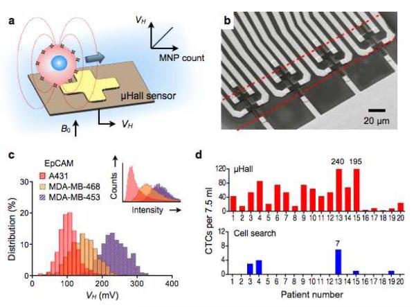
Figure 7. MicroHall (μHall) sensor for single cell detection.
(a) Each cell, targeted with MNPs, generates magnetic fields that are detected by the μHall sensor. The Hall voltage (VH) is proportional to the MNP counts. B0, external magnetic field. (b) The sensing area has a 2 × 4 array of μHall elements. The dotted lines indicate the location of fluidic channel. The sensors are arranged into an overlapping array across the fluidic channel width. (c) The μHall system accurately measured the expression levels of epithelial cell adhesion molecule (EpCAM) in different cell lines; the inset shows the same measurements by flow cytometry. (d) Clinical applications of the μHall system. Circulating tumor cells (CTCs) in patient blood samples (n = 20) were detected using either the μHall system (top) or the clinical gold-standard system, CellSearch (bottom). The μHall enumerated a higher number of CTCs across all patient samples. (Reproduced with permission from ref. 96. Copyright 2012 American Association for the Advancement of Science AAAS.)

