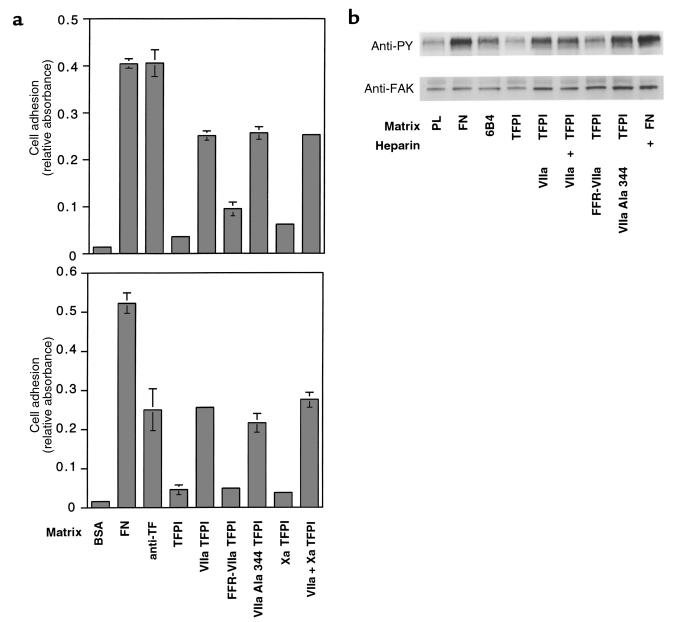
Figure 1.
(a) J82 bladder carcinoma cell adhesion to (top) and migration toward (bottom) the indicated matrices (control [BSA],10 μg/mL fibronectin [FN], 50 μg/mL antibody TF9-6B4 [anti-TF], 100 μg/mL recombinant TFPI-1 [TFPI]) in the presence of 10 nM (adhesion) or 50 nM (migration) recombinant VIIa (VIIa), VIIa covalently active site modified with Phe-Phe-Arg chloromethylketone (FFR-VIIa), VIIa rendered catalytically inactive by catalytic triad Ser 344→Ala mutation (VIIa Ala 344), or factor Xa (Xa). A total of 5 U/mL of heparin was included in all experiments to block proteoglycan-dependent attachment. (b) Phosphorylation of focal adhesion kinase (FAK) in response to adhesion to TFPI-1. Wells were coated with 10 μg/mL poly-L-lysine (PL) or the indicated proteins at concentrations as described for a. Cells were allowed to adhere for 30 minutes in the presence of 5 U/mL heparin where indicated (+), and with the indicated soluble proteins at 10 nM. The top panel shows an anti-phosphotyrosine (anti-PY), and the bottom panel shows an anti-FAK blot of whole-cell lysates.

