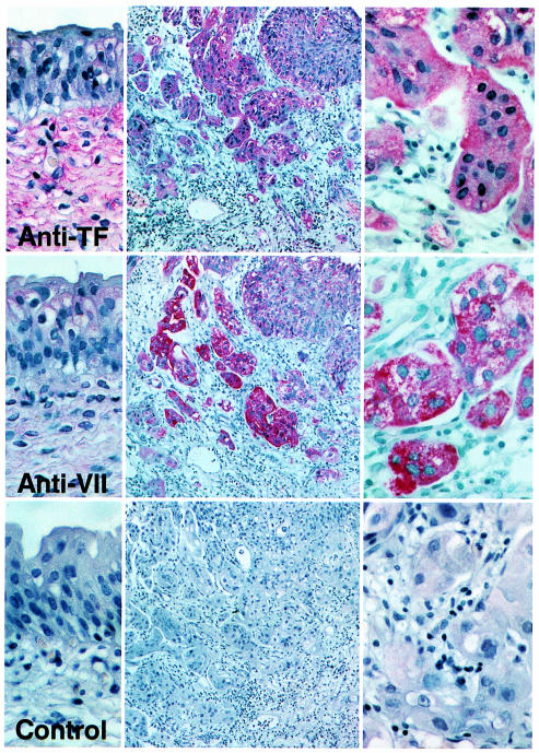
Figure 3.
Immunohistochemical detection of TF and VIIa in invasive bladder carcinoma. The left panels show areas of normal transitional epithelium (×400) adjacent to the invasive areas of the tumor shown as an overview in the middle (×100) and at high magnification in the right panels (×400). The control is staining with anti-TF antibody in the presence of 100 μg/mL soluble TF to demonstrate specificity.

