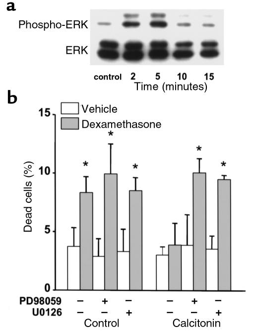
Figure 10.
The antiapoptotic effect of salmon calcitonin involves ERK activation. (a) MLO-Y4 cells were stimulated with 5 ng/mL of sCT for the indicated times. Phosphorylated ERK1/2 and total ERK1/2 were determined by Western blot analysis as described in Methods. (b) Cells were treated for 30 minutes with PD98059 or with UO126, followed by addition of 5 ng/mL of sCT. After 1 hour, 10–6 M dexamethasone was added and cultures were incubated for 6 hours. The percentage of apoptotic cells was determined by trypan blue exclusion, as in Figure 1a. Bars represent the mean ± SD of 3 independent measurements. *P < 0.05 versus control by 1-way ANOVA (Student Newman-Keuls method).

