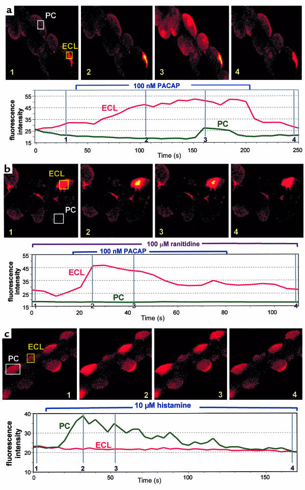
Figure 4.
Effect of PACAP and histamine on superfused Fluo-4–loaded gastric glands using confocal microscopy. (a) In the first sequence, PACAP-38 (100 nM) was added to the perfusate; it can be seen that first the ECL cell [Ca2+]i and then the parietal cell [Ca2+]i is elevated. The position of the images on the trace for [Ca2+]i is presented on the image and in the vertical lines in the lower trace, which shows the calcium level throughout the experiment. The rectangular images represent the regions of the total image that were chosen to generate the scan of the calcium signal. (b) 100 μM ranitidine was added to the perfusate before PACAP addition; although the ECL cell response is retained, the parietal cell response is abolished, as shown in the images and in the trace for [Ca2+]i. (c) Effect of 10 μM histamine on parietal cells [Ca2+]i. The lower panel shows the calcium signal in a parietal cell. No changes were observed in ECL cells with this ligand. Images are representative of at least 4 separate experiments, carried out on different preparations.

