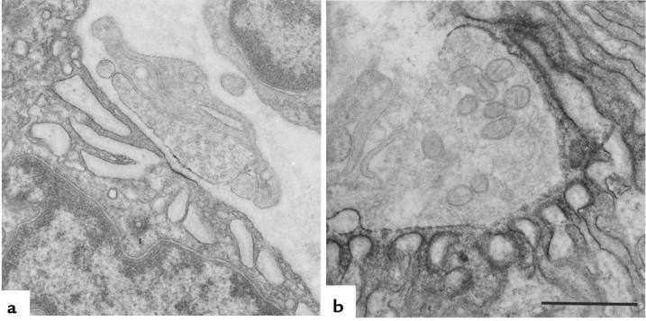
Figure 1.
EP fine structure and localization of AChR at patient (a) and control subject (b) EP regions with peroxidase-labeled α-bgt. Note the small nerve terminal, small postsynaptic region, no openings from the primary synaptic cleft into secondary clefts, and patchy and attenuated reaction for AChR in patient EP.

