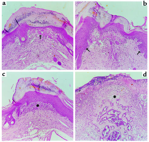
Figure 5.
Histopathological findings on the tail lesions in wild-type animals suffering from EAE. (a) In the earliest stage fibrinoid material (→) appears immediately underneath the thinned epithelium. Inflammatory cells are present in the overlying horny layer. (b) Next, the adjacent epithelium shows irregular hyperplasia (→). (c) At the periphery of the lesion a fibroblastic reaction (*) occurs in the dermis. (d) Irregular outgrowth of osteocartilaginous tissue (*) occurs when the epithelium is completely ulcerated. Hematoxylin and eosin stain; all panels, ×110.

