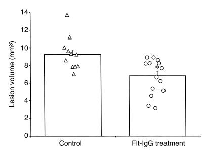
Figure 3.
mFlt-(1–3)-IgG treatment affords long-term tissue salvage as demonstrated by high-resolution anatomical MRI. The size of the infarct can be readily delineated from the coronal MRI sections by measuring the area of the remaining cortex and comparing it with the contralateral hemisphere. Using this approach, the size of the cortical damage was found to be significantly reduced in the mFlt(1–3)-IgG–treated group compared with control group (*P < 0.01).

