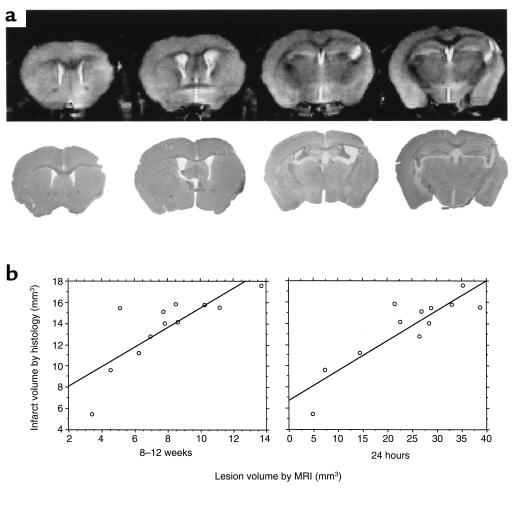
Figure 4.
The histological measure of the infarct size correlates to MRI predictors of infarction. (a) The appearance of the cortical infarction by histology (bottom row), together with the equivalent high resolution MRI (top row) at 8–12 weeks after onset of ischemia. (b) The infarction volume determined by histology was compared with the infarct volume measured with high-resolution, anatomical MRI and the extent of edema as seen from the early T2-weighted MRI. Good correlation between the histology and MRI measurement made at both 8–12 weeks and 24 hours after ischemia/reperfusion was seen.

