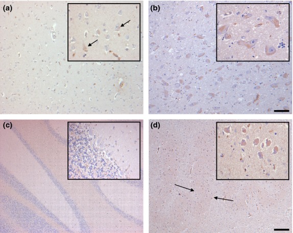
Figure 2.

Distribution of G-CSF IR in the inferior temporal gyrus (a), the basal nucleus of Meynert (b), the cerebellum (c) and the inferior olivary nucleus (d) of controls. Some scattered cortical neurons of the inferior temporal gyrus display faint immunostaining (a, arrows). By contrast, nearly all neurons of the basal nucleus of Meynert are labeled, although IR is only faint (b). In the cerebellum, G-CSF IR is largely absent (c). In the inferior olivary nucleus, all neurons and the neuropil (arrows) are faintly stained (d). Sections are counterstained with hematoxylin. Scale bars: 50 μm (all insets), 100 μm (a,b), and 400 μm (c,d), respectively.
