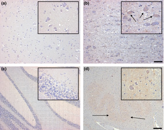
Figure 3.

Distribution of G-CSF R IR in the inferior temporal gyrus (a), the basal nucleus of Meynert (b), the cerebellum (c) and the inferior olivary nucleus (d) of controls. Pyramidal neurons of the inferior temporal gyrus display no immunostaining (a). Scattered neurons of the basal nucleus of Meynert are faintly labeled (b). Within the cerebellar cortex, only weak immunostaining is visible in the granule cell layer (c). In the inferior olivary nucleus, neurons are very weakly labeled, while faint staining is visible in the neuropil (d, arrows). Sections are counterstained with hematoxylin. Scale bar: 50 μm (all insets), 100 μm (a,b) and 400 μm (c,d), respectively.
