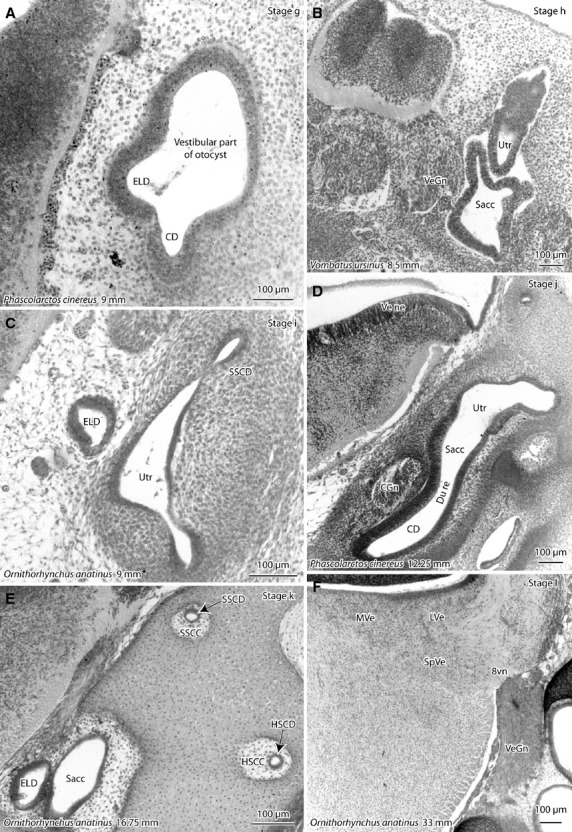
Figure 2.

Photomicrographs of the otocyst/inner ear of developing platypus (Ornithorhynchus anatinus) and marsupials (koala – Phascolarctos cinereus; wombat – Vombatus ursinus) showing stages g–l of vestibular apparatus development (see Table1). Sections are in the frontal plane unless otherwise specified. (A) First appearance of the vestibular portion of the otocyst; (B) first appearance of discrete utricle (Utr), saccule (Sacc) and cochlea (lying out of plane of this sagittal section); (C) first appearance of semicircular ducts (SSCD); (D) narrowing of the ductus reuniens (du re) between the cochlear duct and saccule; (E) semicircular ducts are now surrounded by semicircular canals; (F) differentiation of vestibular nuclei with expansion of neuropil, so that different vestibular nuclei can be distinguished. Species and greatest (body) length of the developing monotreme or marsupial are shown for each image. 8vn, vestibular nerve; CD, cochlear duct; CGn, cochlear ganglion; ELD, endolymphatic duct; HSCC, horizontal semicircular canal; HSCD, horizontal semicircular duct; LVe, lateral vestibular nucleus; MVe, medial vestibular nucleus; SSCC, superior semicircular canal; SSCD, superior semicircular duct; SpVe, spinal vestibular nucleus; VeGn, vestibular ganglion; Ve ne, vestibular neuroepithelium.
