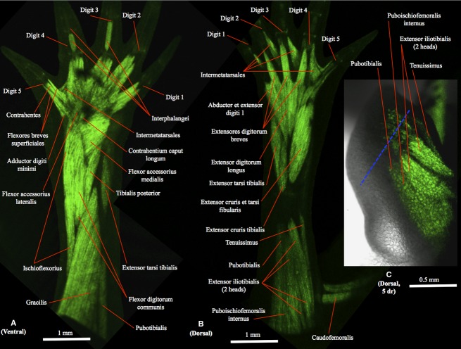
Figure 1.
(A) Ventral (tibial is to the right and distal to the top) and (B) dorsal (tibial is to the left and distal to the top) views of the right hindlimb of GFP-transgenic axolotl CRTD AM125 (10 cm total length) showing a non-amputated limb with a normal muscle configuration, similar to that found in the other non-amputated hindlimbs analyzed for the present study. (C) Dorsal view of the right hindlimb of GFP-transgenic axolotl CRTD AM101 at 5 days of regeneration (dr); tibial is to the left and distal to the top. In this figure and in the next figures the blue dashed line indicates the approximate place of amputation.

