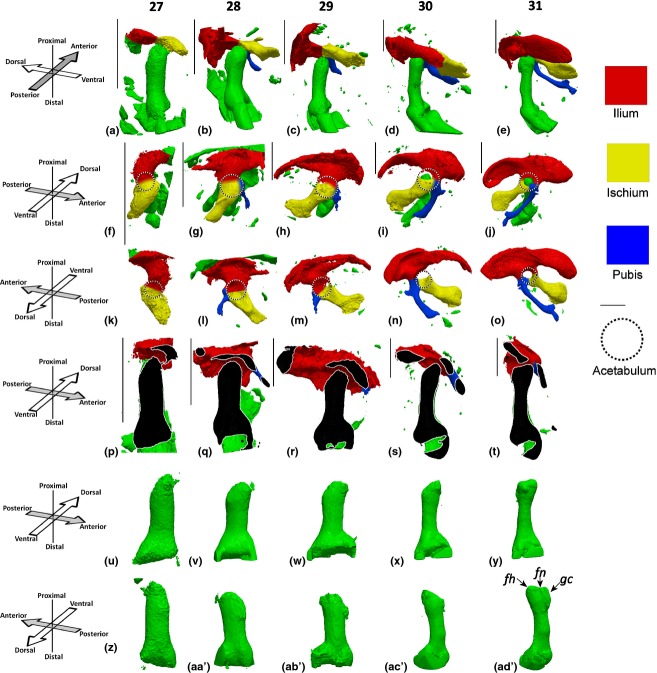
Figure 1.
Pelvic and femoral development between HH27 and HH31. (a–e) Pelvis and femur, posterior aspect of femur; (f–j) pelvis and femur, ventral aspect of pelvis; (k–o) dorsal aspect of pelvis; (p–t) virtual section though the dorsal–ventral plane of the femur, section taken through the femoral head and parallel to the main axis of the femur; (u–y) ventral aspect of the femur, view; (z–ad′) dorsal aspect of the femur. fh, femoral head; fm, femoral neck; gc, greater trochanter. Left limbs shown, orientations for a–e and p–ad′ with respect to femur, orientations for f–o with respect to body axis. Scale bars: 1 mm.

