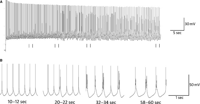
Figure 1.

(A) Example trace recorded from a neuron in the presence of 10 μmol/L TCB‐2, 20 μmol/L DNQX, and 10 μmol/L bicuculline. This cell was held at −42 mV (threshold = −44 mV) for 60 sec by the steady injection of DC current. (B) Expanded sections of A from 10–12, 20–22, 32–34, and 58–60 sec are shown to demonstrate the evolution of rhythmic bursting throughout the trace. Tonic spiking is observed from 0 to 20 sec at which point single spikes are interspersed with doublets as the cell transitions to burst firing. From 30 sec to the end of the trace, the cell fires in a nearly‐exclusive bursting mode. Spike height and burst duration each decrease toward the end of the trace.
