
Figure 1.
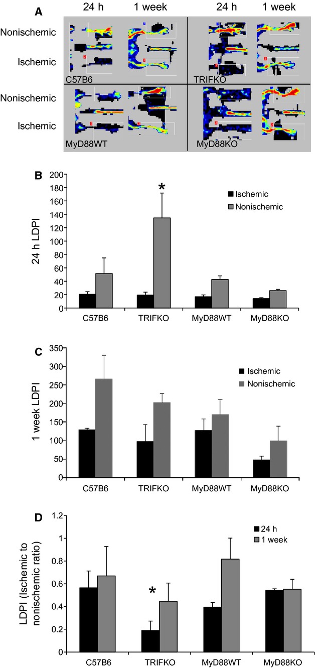
(A) Laser Doppler perfusion images (LDPI) demonstrate the ischemic and nonischemic limbs 24 h and 1 week after right femoral artery ligation. (B) LDPI quantification for each limb after 24 h and 1 week (C). In TRIF KO mice, perfusion to the nonischemic limb is significantly higher than in the other mice (*P =0.02, ANOVA; N =4/group). (D) LDPI ratios of ischemic to nonischemic limb quantified for 24 h and 1 week (*P =0.04, ANOVA; N =4/group).