
Figure 3.
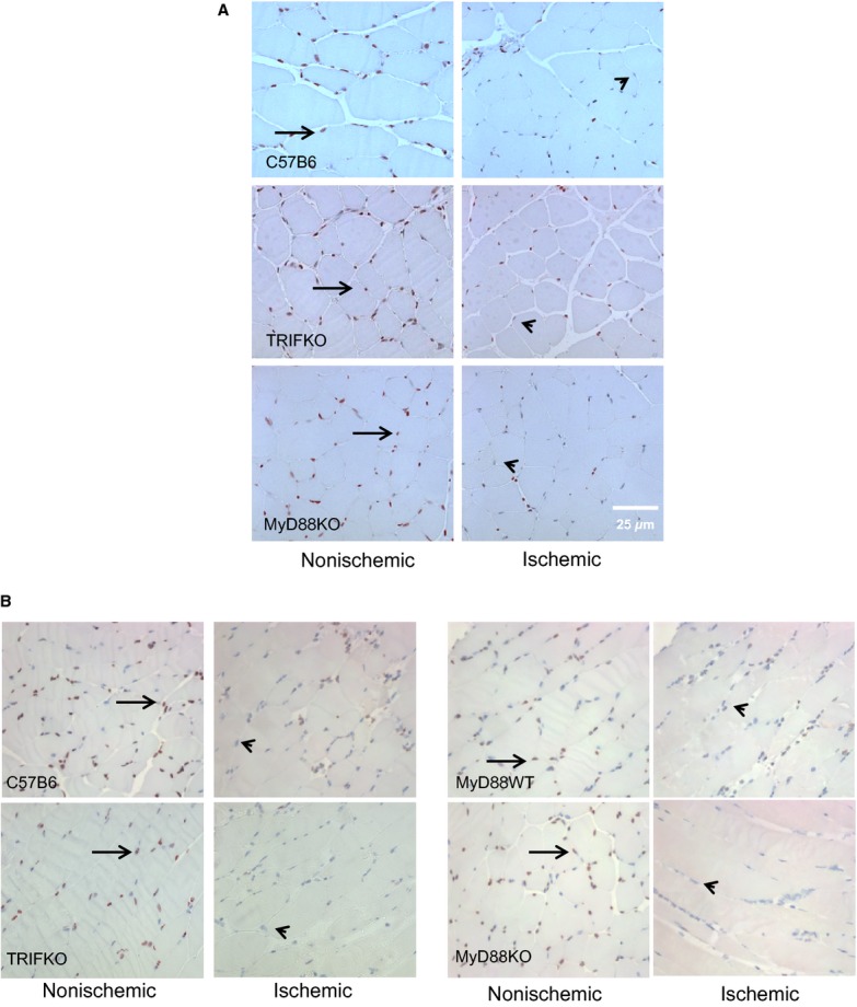
Immunohistochemistry evaluating HMGB1 staining (brown, arrow) in myocyte nuclei (blue, arrowhead) from nonischemic and ischemic limbs 4 (A) and 24 h (B) after femoral artery ligation. The muscle samples were taken from the tibialis anterior compartment.