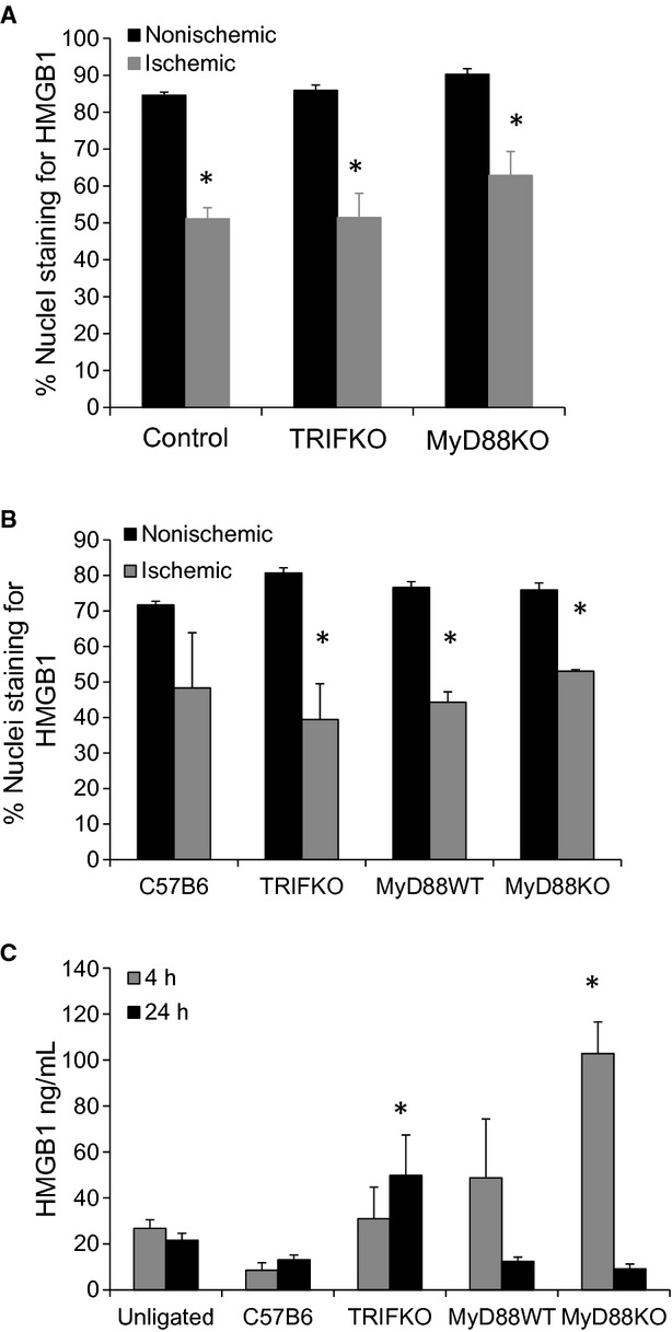
Figure 4.

(A) Nuclear staining of HMGB1 was quantified as percent/total nuclear in control, TRIF KO and MyD88 KO mice 4 h after femoral artery ligation (*P <0.01, t‐test ischemic to nonsichemic; N =3–4/group). (B) The same groups including MyD88 WT mice were evaluated for nuclear staining of HMGB1 24 h after femoral artery ligation (*P <0.01, t‐test ischemic to nonsichemic; N =3–4/group). The muscle samples were taken from the tibialis anterior compartment. (C) Serum HMGB1 was determined 4 and 24 h after femoral artery ligation using ELISA (*P <0.05, ANOVA; N =3–4/group).
