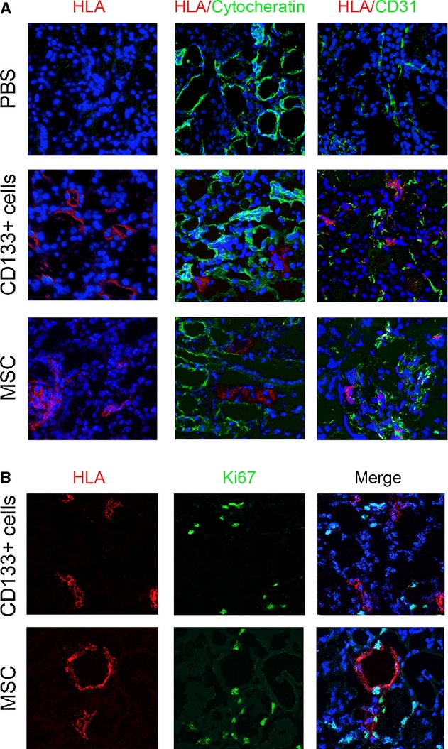
Figure 5.

Detection of CD133+ cells or MSCs within the kidney of AKI mice. (A) Representative confocal micrographs showing the presence of CD133+ cells or MSCs within the kidney of mice with AKI at day 3 after damage as evaluated by HLA Class I (red). Localization of cytokeratin or CD31 positive cells is shown in green. (B) Representative confocal micrographs showing the presence of CD133+ cells or MSCs and of proliferating positive cells within the kidney of mice with AKI at day 3, as evaluated by HLA Class I (red) and Ki67 (green), respectively. Nuclei were counterstained with DAPI (blue). Original magnification 400×.
